
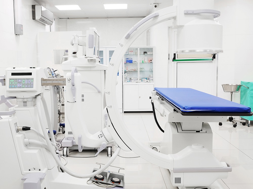
C-Arm fluoroscopy machines serve as versatile instruments in X-ray imaging, catering to diverse patients and medical specialties. Whether you practice in cardiology, orthopedics, or angiography, exploring the role of a C-arm fluoroscopy machines is crucial.
This thorough guide uncovers the details about C-Arm fluoroscopy machines, addressing everything from their definition to their diverse components and uses.
Defining C-Arm Fluoroscopy Machines
A C-Arm fluoroscopy machine utilizes X-rays to generate real-time images of internal bodily structures. Named for its “C” shape, this design facilitates positioning the X-ray source and image intensifier on opposite sides of the patient. This offers exceptional flexibility, allowing healthcare professionals to capture high-quality images from several angles with ease.
Where Are C-Arm Fluoroscopy Machines Used?
C-Arm fluoroscopy machines offer enhanced flexibility, making them ideal for various medical procedures. They are utilized in orthopedics, cardiology, and angiography procedures, as well as therapeutic studies like stent placements and line placements. These systems, characterized by their overhead X-ray image intensification, provide physicians with high-definition views of patients’ anatomical structures. The semi-circular design enables physicians to maneuver the system freely around the patient’s body, capturing images wherever necessary.
What Are the Applications of C-Arm Fluoroscopy Machines?
C-Arm fluoroscopy machines offer versatile applications in several medical procedures. For instance, during complex surgeries requiring needle or stent placement, C-Arm proves invaluable. These machines provide real-time views of vital structures like the gallbladder, liver, and bones, facilitating precise navigation during surgical interventions. With the capability to capture multiple views of the same area, C-Arm fluoroscopy machines enable the creation of detailed 3D models post-surgery. By accurately identifying areas, they enable precise surgical procedures and minimize the risk of damage to the body.
The Parts of a C-Arm Fluoroscopy Machine
- Imaging System
The imaging system of the C-Arm fluoroscopy machine is highly versatile and capable of executing multiple movements within a single procedure. This versatility proves invaluable in a range of surgical fields such as orthopedics, angiography, and cardiology. Despite its compact and lightweight design, the system allows for multiple positions and a wide range of motion. Once mounted, it remains stable throughout the procedure, ensuring zero risk of misalignment.
- X-Ray Generator
The X-ray generator sits within the frame alongside the C-Arm fluoroscopy machine, directly controllable from the workstation unit. Operators can make adjustments to the system’s operation. Even with increased X-ray power, imaging maintains remarkable flexibility, minimizing risks due to reduced exposure times.
- Workstation Unit
The workstation unit serves as the control center for the entire C-Arm operation. It features multiple handles for movements and positioning, switches for power supply and light exposure, a cable hanger, controls for radiographic and fluoroscopic settings, various connecting cables, advanced image enhancement software for noise reduction, contrast and brightness controls, monitors, and a brake pedal.
Contact a Medical Equipment Supplier to Learn More About C-Arm Fluoroscopy Machines
For more information about C-Arm fluoroscopy machines, ask a professional medical equipment supplier.


